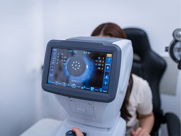
About Advanced Testing and Technology
We believe that great eye care starts with great technology. At our office, we invest in the most advanced tools available to ensure accurate diagnoses, early detection of eye conditions, and personalized care tailored to your needs. Here’s a look at some of the specialized testing and equipment we may use during your visit.
Corneal Topography (Corneal Mapping)
Our corneal mapping system creates a detailed 3D image of the front surface of your eye—the cornea. Since the cornea plays a major role in how your eye focuses light, understanding its shape is key to assessing your vision and eye health.
This non-invasive, painless test helps our team diagnose various corneal conditions, evaluate candidacy for procedures like LASIK, and precisely fit specialty contact lenses. Unlike traditional keratometry, which only samples a few points, this imaging captures thousands of data points across the entire corneal surface in just seconds.
Digital Retinal Imaging & OCT Scanning
We use state-of-the-art imaging tools to get a clear and comprehensive view of your retina and the inner structures of your eye. This allows us to detect signs of serious conditions like glaucoma, diabetic retinopathy, and macular degeneration—even before symptoms appear.
By capturing and storing high-resolution images, we can compare your results over time to monitor for any changes. These scans are fast, non-invasive, and don’t require any special preparation.
Benefits include:
Safe, comfortable, and quick procedure
Ultra-detailed visualization of the retina and sub-layers
Early detection of changes that may impact your vision
No dilation required in most cases
Retinal Photography
With our high-definition retinal camera, we can take images of the back of the eye, including the retina, optic nerve, macula, and blood vessels. This is an essential tool for tracking subtle changes in your eye health—especially if you’re managing ongoing conditions or at risk for retinal disease.
We can also capture images of the front of the eye when needed, allowing us to monitor surface changes over time.
Optical Coherence Tomography (OCT)
Our Heidelberg Spectralis OCT system delivers incredibly precise scans of the retina, allowing us to see cross-sections of the eye’s layers and detect issues that might not be visible with standard tools.
This is particularly useful for evaluating optic nerve health, detecting retinal swelling or bleeding, and diagnosing conditions like macular degeneration and glaucoma. With advanced tracking features, the system can rescan the exact same area at future visits to detect even the smallest changes—sometimes as little as 3 microns. We also coordinate closely with retinal specialists and can share these detailed scans directly with their offices, helping streamline your care.
Visual Field Testing
This test maps your peripheral (side) vision and helps identify blind spots or visual field loss—often associated with glaucoma, stroke, or neurological conditions. The test compares your responses to normal values for your age group and provides a baseline for monitoring progression over time.
Visual field testing can help detect or monitor:
Glaucoma and ocular hypertension
Diabetic retinopathy
Macular conditions (edema, holes, degeneration)
Vascular changes or injury
Stroke and traumatic brain injury
Retinal scars or pigment changes
Committed to Your Eye Health
Our practice is proud to combine compassionate care with the latest in diagnostic innovation. With every test, scan, and image, our goal is to protect your vision and give you the clearest possible view of the world.


